
| [1] |
Sung H, Ferlay J, Siegel R L, et al. Global cancer statistics 2020: GLOBOCAN estimates of incidence and mortality worldwide for 36 cancers in 185 countries. CA Cancer J. Clin., 2021, 71 (3): 209–249. doi: 10.3322/caac.21660
|
| [2] |
Gram I T, Park S Y, Wilkens L R, et al. Smoking-related risks of colorectal cancer by anatomical subsite and sex. Am. J. Epidemiol., 2020, 189 (6): 543–553. doi: 10.1093/aje/kwaa005
|
| [3] |
Akter S, Islam Z, Mizoue T, et al. Smoking and colorectal cancer: A pooled analysis of 10 population-based cohort studies in Japan. Int. J. Cancer, 2021, 148 (3): 654–664. doi: 10.1002/ijc.33248
|
| [4] |
Tarasiuk A, Mosińska P, Fichna J. The mechanisms linking obesity to colon cancer: An overview. Obes. Res. Clin. Pract., 2018, 12 (3): 251–259. doi: 10.1016/j.orcp.2018.01.005
|
| [5] |
Tverdal A, Høiseth G, Magnus P, et al. Alcohol consumption, HDL-cholesterol and incidence of colon and rectal cancer: a prospective cohort study including 250,010 participants. Alcohol Alcoholism, 2021, 56 (6): 718–725. doi: 10.1093/alcalc/agab007
|
| [6] |
Lynch H T, Snyder C L, Shaw T G, et al. Milestones of Lynch syndrome: 1895-2015. Nat. Rev. Cancer, 2015, 15 (3): 181–194. doi: 10.1038/nrc3878
|
| [7] |
Li J Y, Wang R, Zhou X, et al. Genomic and transcriptomic profiling of carcinogenesis in patients with familial adenomatous polyposis. Gut, 2020, 69 (7): 1283–1293. doi: 10.1136/gutjnl-2019-319438
|
| [8] |
Jasperson K W, Tuohy T M, Neklason D W, et al. Hereditary and familial colon cancer. Gastroenterology, 2010, 138 (6): 2044–2058. doi: 10.1053/j.gastro.2010.01.054
|
| [9] |
Bian J, Dannappel M, Wan C, et al. Transcriptional regulation of Wnt/β-catenin pathway in colorectal cancer. Cells, 2020, 9 (9): 2125. doi: 10.3390/cells9092125
|
| [10] |
Yang L, Lin C, Jin C, et al. lncRNA-dependent mechanisms of androgen-receptor-regulated gene activation programs. Nature, 2013, 500 (7464): 598–602. doi: 10.1038/nature12451
|
| [11] |
Li G, Kryczek I, Nam J, et al. LIMIT is an immunogenic lncRNA in cancer immunity and immunotherapy. Nat. Cell Biol., 2021, 23 (5): 526–537. doi: 10.1038/s41556-021-00672-3
|
| [12] |
Huang D, Chen J, Yang L, et al. NKILA lncRNA promotes tumor immune evasion by sensitizing T cells to activation-induced cell death. Nat. Immunol., 2018, 19 (10): 1112–1125. doi: 10.1038/s41590-018-0207-y
|
| [13] |
Jiang W X, Pan S Y, Chen X, et al. The role of lncRNAs and circRNAs in the PD-1/PD-L1 pathway in cancer immunotherapy. Mol. Cancer, 2021, 20 (1): 116. doi: 10.1186/s12943-021-01406-7
|
| [14] |
Xu H, Jiang Y, Xu X, et al. Inducible degradation of lncRNA Sros1 promotes IFN-γ-mediated activation of innate immune responses by stabilizing Stat1 mRNA. Nat. Immunol., 2019, 20 (12): 1621–1630. doi: 10.1038/s41590-019-0542-7
|
| [15] |
Mills C D, Lenz L L, Harris R A. A breakthrough: Macrophage-directed cancer immunotherapy. Cancer Res., 2016, 76 (3): 513–516. doi: 10.1158/0008-5472.CAN-15-1737
|
| [16] |
Choo Y W, Kang M, Kim H Y, et al. M1 macrophage-derived nanovesicles potentiate the anticancer efficacy of immune checkpoint inhibitors. ACS Nano, 2018, 12 (9): 8977–8993. doi: 10.1021/acsnano.8b02446
|
| [17] |
Li M, Sun X, Zhao J, et al. CCL5 deficiency promotes liver repair by improving inflammation resolution and liver regeneration through M2 macrophage polarization. Cell. Mol. Immunol., 2020, 17 (7): 753–764. doi: 10.1038/s41423-019-0279-0
|
| [18] |
Vogel D Y S, Glim J E, Stavenuiter A W D, et al. Human macrophage polarization in vitro: Maturation and activation methods compared. Immunobiology, 2014, 219 (9): 695–703. doi: 10.1016/j.imbio.2014.05.002
|
| [19] |
Lan J Q, Sun L, Xu F, et al. M2 macrophage-derived exosomes promote cell migration and invasion in colon cancer. Cancer Res., 2019, 79 (1): 146–158. doi: 10.1158/0008-5472.CAN-18-0014
|
| [20] |
Yamaguchi T, Fushida S, Yamamoto Y, et al. Tumor-associated macrophages of the M2 phenotype contribute to progression in gastric cancer with peritoneal dissemination. Gastric Cancer, 2016, 19 (4): 1052–1065. doi: 10.1007/s10120-015-0579-8
|
| [21] |
Sharma A, Seow J J W, Dutertre C A, et al. Onco-fetal reprogramming of endothelial cells drives immunosuppressive macrophages in hepatocellular carcinoma. Cell, 2020, 183 (2): 377–394.e21. doi: 10.1016/j.cell.2020.08.040
|
| [22] |
Cassetta L, Fragkogianni S, Sims A H, et al. Human tumor-associated macrophage and monocyte transcriptional landscapes reveal cancer-specific reprogramming, biomarkers, and therapeutic targets. Cancer Cell, 2019, 35 (4): 588–602.e10. doi: 10.1016/j.ccell.2019.02.009
|
| [23] |
Morrissey SM, Zhang F, Ding C L, et al. Tumor-derived exosomes drive immunosuppressive macrophages in a pre-metastatic niche through glycolytic dominant metabolic reprogramming. Cell Metab., 2021, 33 (10): 2040–2058.e10. doi: 10.1016/j.cmet.2021.09.002
|
| [24] |
Franklin R A, Liao W, Sarkar A, et al. The cellular and molecular origin of tumor-associated macrophages. Science, 2014, 344 (6186): 921–925. doi: 10.1126/science.1252510
|
| [25] |
Zigmond R E, Echevarria F D. Macrophage biology in the peripheral nervous system after injury. Prog. Neurobiol., 2019, 173: 102–121. doi: 10.1016/j.pneurobio.2018.12.001
|
| [26] |
Dan H, Liu S, Liu J, et al. RACK1 promotes cancer progression by increasing the M2/M1 macrophage ratio via the NF-κB pathway in oral squamous cell carcinoma. Mol. Oncol., 2020, 14 (4): 795–807. doi: 10.1002/1878-0261.12644
|
| [27] |
Boimel P J, Smirnova T, Zhou Z N, et al. Contribution of CXCL12 secretion to invasion of breast cancer cells. Breast Cancer Res., 2012, 14 (1): R23. doi: 10.1186/bcr3108
|
| [28] |
Newman A M, Steen C B, Liu C L, et al. Determining cell type abundance and expression from bulk tissues with digital cytometry. Nat. Biotechnol., 2019, 37 (7): 773–782. doi: 10.1038/s41587-019-0114-2
|
| [29] |
Love M I, Huber W, Anders S. Moderated estimation of fold change and dispersion for RNA-seq data with DESeq2. Genome Biol., 2014, 15 (12): 550. doi: 10.1186/s13059-014-0550-8
|
| [30] |
Volders P J, Anckaert J, Verheggen K, et al. LNCipedia 5: towards a reference set of human long non-coding RNAs. Nucleic Acids Res., 2019, 47 (D1): D135–D139. doi: 10.1093/nar/gky1031
|
| [31] |
Subramanian A, Tamayo P, Mootha V K, et al. Gene set enrichment analysis: A knowledge-based approach for interpreting genome-wide expression profiles. Proc. Natl. Acad. Sci. U.S.A., 2005, 102 (43): 15545–15550. doi: 10.1073/pnas.0506580102
|
| [32] |
Mootha V K, Lindgren C M, Eriksson K F, et al. PGC-1α-responsive genes involved in oxidative phosphorylation are coordinately downregulated in human diabetes. Nat. Genet., 2003, 34 (3): 267–273. doi: 10.1038/ng1180
|
| [33] |
Raudvere U, Kolberg L, Kuzmin I, et al. g:Profiler: a web server for functional enrichment analysis and conversions of gene lists (2019 update). Nucleic Acids Res., 2019, 47 (W1): W191–W198. doi: 10.1093/nar/gkz369
|
| [34] |
Mayakonda A, Lin D C, Assenov Y, et al. Maftools: efficient and comprehensive analysis of somatic variants in cancer. Genome Res., 2018, 28 (11): 1747–1756. doi: 10.1101/gr.239244.118
|
| [35] |
Diao Z L, Han Y X, Chen Y Q, et al. The clinical utility of microsatellite instability in colorectal cancer. Crit. Rev. Oncol. Hematol., 2021, 157: 103171. doi: 10.1016/j.critrevonc.2020.103171
|
| [36] |
Schrock A B, Ouyang C, Sandhu J, et al. Tumor mutational burden is predictive of response to immune checkpoint inhibitors in MSI-high metastatic colorectal cancer. Ann. Oncol., 2019, 30 (7): 1096–1103. doi: 10.1093/annonc/mdz134
|
| [37] |
Ganesh K, Stadler Z K, Cercek A, et al. Immunotherapy in colorectal cancer: Rationale, challenges and potential. Nat. Rev. Gastroenterol. Hepatol., 2019, 16 (6): 361–375. doi: 10.1038/s41575-019-0126-x
|
| [38] |
Chida K, Kawazoe A, Kawazu M, et al. A low tumor mutational burden and PTEN mutations are predictors of a negative response to PD-1 blockade in MSI-H/dMMR gastrointestinal tumors. Clin. Cancer Res., 2021, 27 (13): 3714–3724. doi: 10.1158/1078-0432.CCR-21-0401
|
| [39] |
Chalmers Z R, Connelly C F, Fabrizio D, et al. Analysis of 100,000 human cancer genomes reveals the landscape of tumor mutational burden. Genome Med., 2017, 9 (1): 34. doi: 10.1186/s13073-017-0424-2
|
| [40] |
Jardim D L, Goodman A, de Melo Gagliato D, et al. The challenges of tumor mutational burden as an immunotherapy biomarker. Cancer Cell, 2021, 39 (2): 154–173. doi: 10.1016/j.ccell.2020.10.001
|
| [41] |
Zhou M, Zhang Z C, Bao S Q, et al. Computational recognition of lncRNA signature of tumor-infiltrating B lymphocytes with potential implications in prognosis and immunotherapy of bladder cancer. Brief. Bioinform., 2021, 22 (3): bbaa047. doi: 10.1093/bib/bbaa047
|
| [42] |
Xia Y, Rao L, Yao H, et al. Engineering macrophages for cancer immunotherapy and drug delivery. Adv. Mater., 2020, 32 (40): 2002054. doi: 10.1002/adma.202002054
|
| [43] |
Mohapatra S, Pioppini C, Ozpolat B, et al. Non-coding RNAs regulation of macrophage polarization in cancer. Mol. Cancer, 2021, 20 (1): 24. doi: 10.1186/s12943-021-01313-x
|
| [44] |
Gunassekaran G R, Poongkavithai Vadevoo S M, Baek M C, et al. M1 macrophage exosomes engineered to foster M1 polarization and target the IL-4 receptor inhibit tumor growth by reprogramming tumor-associated macrophages into M1-like macrophages. Biomaterials, 2021, 278: 121137. doi: 10.1016/j.biomaterials.2021.121137
|
| [45] |
Li C, Hu G, Wei B, et al. lncRNA LINC01494 promotes proliferation, migration and invasion in glioma through miR-122-5p/CCNG1 axis. OncoTargets Ther., 2019, 12: 7655–7662. doi: 10.2147/OTT.S213345
|
| [46] |
Bao Z, Yang Z, Huang Z, et al. LncRNADisease 2.0: an updated database of long non-coding RNA-associated diseases. Nucleic Acids Res., 2019, 47 (D1): D1034–D1037. doi: 10.1093/nar/gky905
|
| [47] |
Liang Z X, Liu H S, Wang F W, et al. LncRNA RPPH1 promotes colorectal cancer metastasis by interacting with TUBB3 and by promoting exosomes-mediated macrophage M2 polarization. Cell Death Dis., 2019, 10 (11): 829. doi: 10.1038/s41419-019-2077-0
|
| [48] |
Xu Z J, Chen Y, Ma L, et al. Role of exosomal non-coding RNAs from tumor cells and tumor-associated macrophages in the tumor microenvironment. Mol. Ther., 2022, 30 (10): 3133–3154. doi: 10.1016/j.ymthe.2022.01.046
|
| [49] |
Zhou R, Zhang J W, Zeng D Q, et al. Immune cell infiltration as a biomarker for the diagnosis and prognosis of stage I–III colon cancer. Cancer Immunol. Immunother., 2019, 68 (3): 433–442. doi: 10.1007/s00262-018-2289-7
|
| [50] |
Galon J, Costes A, Sanchez-Cabo F, et al. Type, density, and location of immune cells within human colorectal tumors predict clinical outcome. Science, 2006, 313 (5795): 1960–1964. doi: 10.1126/science.1129139
|
| [51] |
Bagchi S, Yuan R, Engleman E G. Immune checkpoint inhibitors for the treatment of cancer: Clinical impact and mechanisms of response and resistance. Annu. Rev. Pathol., 2021, 16: 223–249. doi: 10.1146/annurev-pathol-042020-042741
|
| [52] |
Popat S, Hubner R, Houlston R S. Systematic review of microsatellite instability and colorectal cancer prognosis. J. Clin. Oncol., 2005, 23 (3): 609–618. doi: 10.1200/JCO.2005.01.086
|
| [53] |
Gryfe R, Kim H, Hsieh E T K, et al. Tumor microsatellite instability and clinical outcome in young patients with colorectal cancer. N. Engl. J. Med, 2000, 342 (2): 69–77. doi: 10.1056/NEJM200001133420201
|
| [54] |
Vilar E, Gruber S B. Microsatellite instability in colorectal cancer—the stable evidence. Nat. Rev. Clin. Oncol., 2010, 7 (3): 153–162. doi: 10.1038/nrclinonc.2009.237
|
| [55] |
Bellone S, Roque D M, Siegel E R, et al. A phase II evaluation of pembrolizumab in recurrent microsatellite instability-high (MSI-H) endometrial cancer patients with Lynch-like versus MLH-1 methylated characteristics (NCT02899793). Ann. Oncol., 2021, 32 (8): 1045–1046. doi: 10.1016/j.annonc.2021.04.013
|
| [56] |
Puliga E, Corso S, Pietrantonio F, et al. Microsatellite instability in Gastric Cancer: Between lights and shadows. Cancer Treat. Rev., 2021, 95: 102175. doi: 10.1016/j.ctrv.2021.102175
|
| [57] |
Pal T, Permuth-Wey J, Kumar A, et al. Systematic review and meta-analysis of ovarian cancers: Estimation of microsatellite-high frequency and characterization of mismatch repair deficient tumor histology. Clin. Cancer Res., 2008, 14 (21): 6847–6854. doi: 10.1158/1078-0432.CCR-08-1387
|
| [58] |
Gstalder C, Liu D, Miao D, et al. Inactivation of Fbxw7 impairs dsRNA sensing and confers resistance to PD-1 blockade. Cancer Discov., 2020, 10 (9): 1296–1311. doi: 10.1158/2159-8290.CD-19-1416
|
| [59] |
Qi J, Sun H, Zhang Y, et al. Single-cell and spatial analysis reveal interaction of FAP+ fibroblasts and SPP1+ macrophages in colorectal cancer. Nat. Commun., 2022, 13 (1): 1742. doi: 10.1038/s41467-022-29366-6
|
| [60] |
Zhou L, Wang M, Guo H, et al. Integrated analysis highlights the immunosuppressive role of TREM2+ macrophages in hepatocellular carcinoma. Front. Immunol., 2022, 13: 848367. doi: 10.3389/fimmu.2022.848367
|
| [61] |
Katzenelenbogen Y, Sheban F, Yalin A, et al. Coupled scRNA-seq and intracellular protein activity reveal an immunosuppressive role of TREM2 in cancer. Cell, 2020, 182 (4): 872–885.e19. doi: 10.1016/j.cell.2020.06.032
|
| [62] |
Bugatti M, Bergamini M, Missale F, et al. A population of TIM4+FOLR2+ macrophages localized in tertiary lymphoid structures correlates to an active immune infiltrate across several cancer types. Cancer Immunol. Res., 2022, 10 (11): 1340–1353. doi: 10.1158/2326-6066.CIR-22-0271
|
| [63] |
Nalio Ramos R, Missolo-Koussou Y, Gerber-Ferder Y, et al. Tissue-resident FOLR2+ macrophages associate with CD8+ T cell infiltration in human breast cancer. Cell, 2022, 185 (7): 1189–1207.e25. doi: 10.1016/j.cell.2022.02.021
|
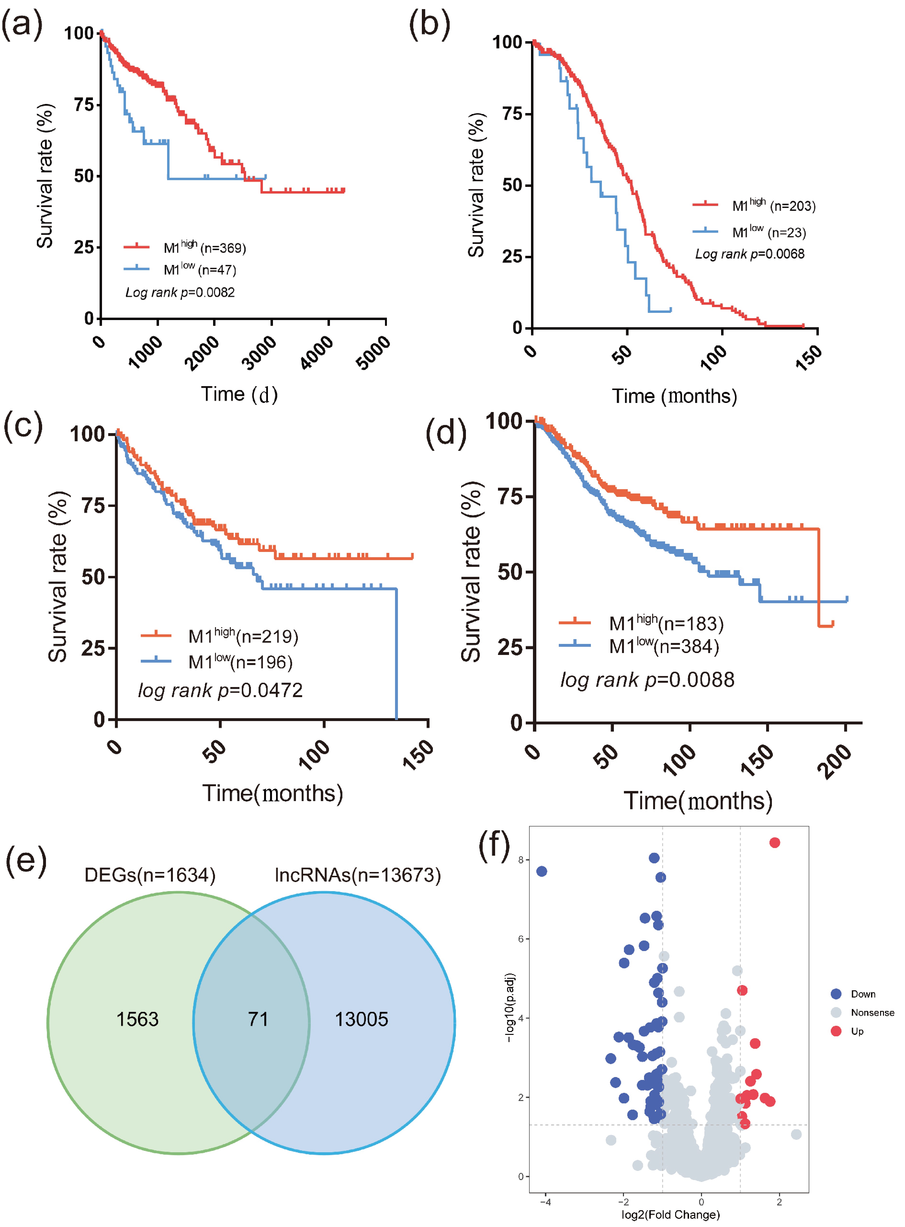
Figure 1. M1-Mφ predicts the clinical prognosis of colon cancer patients and DElncRNA detection. (a–d) Kaplan‒Meier curve of TCGA-COAD dataset, GSE14333, integrated dataset of GSE17536 and GSE17538, GSE39582, respectively, stratified by M1 macrophage proportion. (e) Venn diagram shows DElncRNAs through intersecting DEGs (n=1634) with the lncRNA list (n=13076), 71 DElncRNAs containing 58 downregulated lncRNAs and 13 upregulated lncRNAs. (f) Volcano plot of DElncRNAs, with upregulated lncRNAs shown in red (n=13), downregulated lncRNAs shown in blue (n=58, Wilcoxon rank sum test) and nonsense lncRNAs shown in gray (n=1180, Wilcoxon rank sum test).

Figure 2. The constructed lncRNA signature is closely associated with clinical outcomes. (a–g) Kaplan‒Meier curve of 7 survival-related lncRNAs. (a) AADACL2-AS (log rank p=0.0037); (b) EVX1-AS (log rank p =0.0338); (c) KIF25-AS1 (log rank p =0.0013); (d) LINC00871 (log rank p =0.0013); (e) LINC01450 (log rank p =0.0117); (f) LINC01494 (log rank p =0.0011); (g) ZHDDC20-IT1 (log rank p = 0.0043). (h) Kaplan‒Meier curve of the TCGA-COAD cohort (left, log rank p=0.0007) and an independent validation dataset GSE17537 (log rank p=0.0106). (i) Box plot of the M1 macrophage infiltration proportion in the lncRNAhigh and lncRNAlow groups (p<0.0001, Mann‒Whitney test, data are represented as the mean ± SD). (j) Forest plot of the multivariate Cox regression analysis of the TCGA-COAD cohort (left) and the independent validation cohort GSE17537 (right). (k) Box plot of the lncRNA score of different COAD tumor stages (Mann‒Whitney test, compared to stage i COAD tumors, data are represented as the mean ± SD).
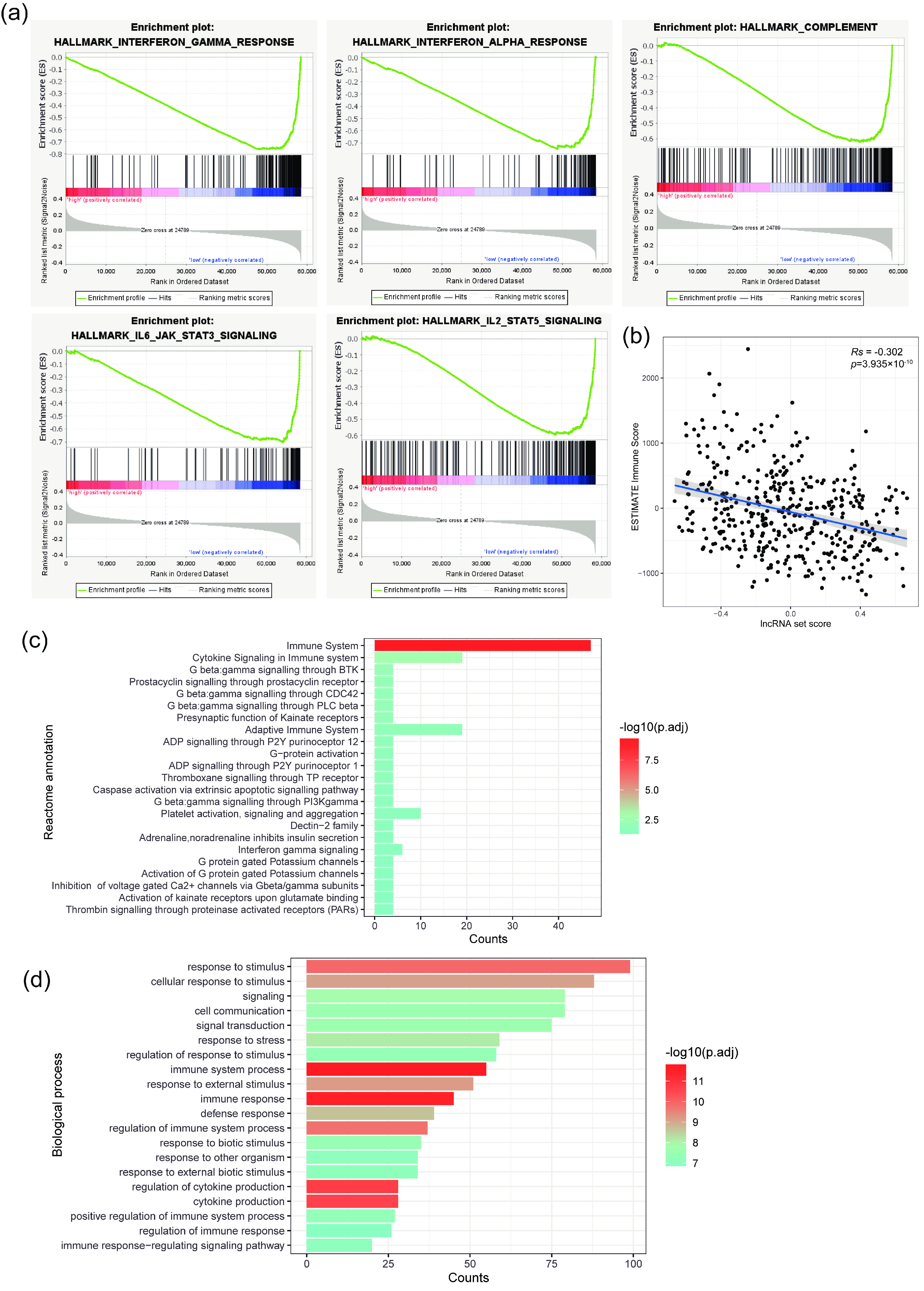
Figure 3. The tumor immune microenvironment is negatively correlated with the lncRNA signature. (a) GSEA plots of differentially enriched cancer hallmark pathways between the lncRNA scorehigh and lncRNA scorelow cohorts. (b) Spearman’s correlation between lncRNA score and ESTIMATE immune score (Rs=−0.302, p=3.935×10⁻¹⁰). (c) Reactome enrichment plot of mRNAs that negatively correlated with the lncRNA set (n=23, Fisher’s exact test). (d) GO:BP enrichment plot of mRNAs that negatively correlated with the lncRNA signature. The top 20 enriched biological processes are displayed (Fisher’s exact test).

Figure 4. Antigen-presenting processes were downregulated in the lncRNA scorehigh group. (a) Spearman correlation between lncRNA score and antigen presenting and processing score (Rs=−0.307, p=2.054×10⁻¹⁰). (b) Box plot of differences in chemotaxis score between the high lncRNA group and the low lncRNA group (**: p<0.05, ***:p<0.0001, Mann‒Whitney test, data are represented as the mean ± SD). (c) Spearman correlation of lncRNA score and genes that positively regulate antigen processing and presentation. (d) Spearman correlation of lncRNA score and genes that positively regulate chemotaxis.
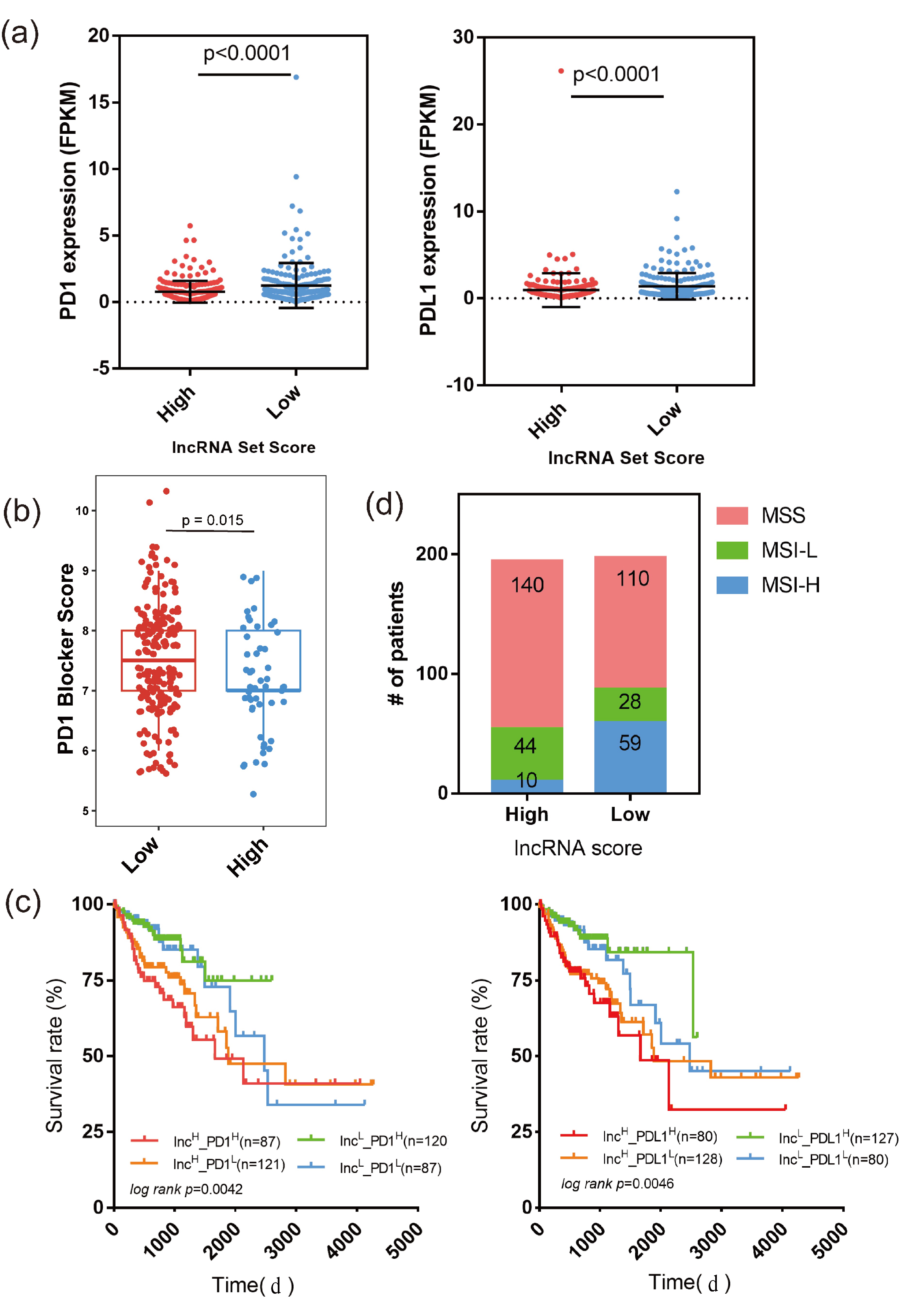
Figure 5. LncRNA signature predicts ICI therapy outcomes. (a) Swarm diagram shows the differences in PD1 (left) and PD-L1 (right) between the lncRNA scorehigh and lncRNA scorelow groups (data are represented as the mean ± SD, Mann‒Whitney test). (b) Predictive value of PD1 blocker efficiency between the lncRNA set scorehigh and scorelow groups (p=0.015, Student’s t test) (c) Kaplan‒Meier curve of four patient groups stratified by lncRNA score and PD1 expression (left) and lncRNA score and PD-L1 expression (right). (d) Stacked histograms of MSS, MSI-L and MSI-H patient numbers in the lncRNA scorehigh and lncRNA scorelow groups (p<0.05, chi-square test).

Figure 6. Relationship between tumor mutation burden and lncRNA signature. (a) Swarm diagram shows the differences in TMB between the lncRNA scorehigh and lncRNA scorelow groups (data are represented as the mean ± SD, Mann‒Whitney test). (b) Waterfall plot shows the top 30 mutated genes in the lncRNA scorehigh (upper panel) and lncRNA scorelow groups (lower panel). Genes in black suggest shared mutated genes across the lncRNA scorehigh and lncRNA scorelow groups. Genes in red were highly mutated in the lncRNA scorehigh group. Genes in cyan were highly mutated in the lncRNA scorelow group. (c) Comparing MSI-related gene mutations between the lncRNA scorehigh and lncRNA scorelow groups.
| [1] |
Sung H, Ferlay J, Siegel R L, et al. Global cancer statistics 2020: GLOBOCAN estimates of incidence and mortality worldwide for 36 cancers in 185 countries. CA Cancer J. Clin., 2021, 71 (3): 209–249. doi: 10.3322/caac.21660
|
| [2] |
Gram I T, Park S Y, Wilkens L R, et al. Smoking-related risks of colorectal cancer by anatomical subsite and sex. Am. J. Epidemiol., 2020, 189 (6): 543–553. doi: 10.1093/aje/kwaa005
|
| [3] |
Akter S, Islam Z, Mizoue T, et al. Smoking and colorectal cancer: A pooled analysis of 10 population-based cohort studies in Japan. Int. J. Cancer, 2021, 148 (3): 654–664. doi: 10.1002/ijc.33248
|
| [4] |
Tarasiuk A, Mosińska P, Fichna J. The mechanisms linking obesity to colon cancer: An overview. Obes. Res. Clin. Pract., 2018, 12 (3): 251–259. doi: 10.1016/j.orcp.2018.01.005
|
| [5] |
Tverdal A, Høiseth G, Magnus P, et al. Alcohol consumption, HDL-cholesterol and incidence of colon and rectal cancer: a prospective cohort study including 250,010 participants. Alcohol Alcoholism, 2021, 56 (6): 718–725. doi: 10.1093/alcalc/agab007
|
| [6] |
Lynch H T, Snyder C L, Shaw T G, et al. Milestones of Lynch syndrome: 1895-2015. Nat. Rev. Cancer, 2015, 15 (3): 181–194. doi: 10.1038/nrc3878
|
| [7] |
Li J Y, Wang R, Zhou X, et al. Genomic and transcriptomic profiling of carcinogenesis in patients with familial adenomatous polyposis. Gut, 2020, 69 (7): 1283–1293. doi: 10.1136/gutjnl-2019-319438
|
| [8] |
Jasperson K W, Tuohy T M, Neklason D W, et al. Hereditary and familial colon cancer. Gastroenterology, 2010, 138 (6): 2044–2058. doi: 10.1053/j.gastro.2010.01.054
|
| [9] |
Bian J, Dannappel M, Wan C, et al. Transcriptional regulation of Wnt/β-catenin pathway in colorectal cancer. Cells, 2020, 9 (9): 2125. doi: 10.3390/cells9092125
|
| [10] |
Yang L, Lin C, Jin C, et al. lncRNA-dependent mechanisms of androgen-receptor-regulated gene activation programs. Nature, 2013, 500 (7464): 598–602. doi: 10.1038/nature12451
|
| [11] |
Li G, Kryczek I, Nam J, et al. LIMIT is an immunogenic lncRNA in cancer immunity and immunotherapy. Nat. Cell Biol., 2021, 23 (5): 526–537. doi: 10.1038/s41556-021-00672-3
|
| [12] |
Huang D, Chen J, Yang L, et al. NKILA lncRNA promotes tumor immune evasion by sensitizing T cells to activation-induced cell death. Nat. Immunol., 2018, 19 (10): 1112–1125. doi: 10.1038/s41590-018-0207-y
|
| [13] |
Jiang W X, Pan S Y, Chen X, et al. The role of lncRNAs and circRNAs in the PD-1/PD-L1 pathway in cancer immunotherapy. Mol. Cancer, 2021, 20 (1): 116. doi: 10.1186/s12943-021-01406-7
|
| [14] |
Xu H, Jiang Y, Xu X, et al. Inducible degradation of lncRNA Sros1 promotes IFN-γ-mediated activation of innate immune responses by stabilizing Stat1 mRNA. Nat. Immunol., 2019, 20 (12): 1621–1630. doi: 10.1038/s41590-019-0542-7
|
| [15] |
Mills C D, Lenz L L, Harris R A. A breakthrough: Macrophage-directed cancer immunotherapy. Cancer Res., 2016, 76 (3): 513–516. doi: 10.1158/0008-5472.CAN-15-1737
|
| [16] |
Choo Y W, Kang M, Kim H Y, et al. M1 macrophage-derived nanovesicles potentiate the anticancer efficacy of immune checkpoint inhibitors. ACS Nano, 2018, 12 (9): 8977–8993. doi: 10.1021/acsnano.8b02446
|
| [17] |
Li M, Sun X, Zhao J, et al. CCL5 deficiency promotes liver repair by improving inflammation resolution and liver regeneration through M2 macrophage polarization. Cell. Mol. Immunol., 2020, 17 (7): 753–764. doi: 10.1038/s41423-019-0279-0
|
| [18] |
Vogel D Y S, Glim J E, Stavenuiter A W D, et al. Human macrophage polarization in vitro: Maturation and activation methods compared. Immunobiology, 2014, 219 (9): 695–703. doi: 10.1016/j.imbio.2014.05.002
|
| [19] |
Lan J Q, Sun L, Xu F, et al. M2 macrophage-derived exosomes promote cell migration and invasion in colon cancer. Cancer Res., 2019, 79 (1): 146–158. doi: 10.1158/0008-5472.CAN-18-0014
|
| [20] |
Yamaguchi T, Fushida S, Yamamoto Y, et al. Tumor-associated macrophages of the M2 phenotype contribute to progression in gastric cancer with peritoneal dissemination. Gastric Cancer, 2016, 19 (4): 1052–1065. doi: 10.1007/s10120-015-0579-8
|
| [21] |
Sharma A, Seow J J W, Dutertre C A, et al. Onco-fetal reprogramming of endothelial cells drives immunosuppressive macrophages in hepatocellular carcinoma. Cell, 2020, 183 (2): 377–394.e21. doi: 10.1016/j.cell.2020.08.040
|
| [22] |
Cassetta L, Fragkogianni S, Sims A H, et al. Human tumor-associated macrophage and monocyte transcriptional landscapes reveal cancer-specific reprogramming, biomarkers, and therapeutic targets. Cancer Cell, 2019, 35 (4): 588–602.e10. doi: 10.1016/j.ccell.2019.02.009
|
| [23] |
Morrissey SM, Zhang F, Ding C L, et al. Tumor-derived exosomes drive immunosuppressive macrophages in a pre-metastatic niche through glycolytic dominant metabolic reprogramming. Cell Metab., 2021, 33 (10): 2040–2058.e10. doi: 10.1016/j.cmet.2021.09.002
|
| [24] |
Franklin R A, Liao W, Sarkar A, et al. The cellular and molecular origin of tumor-associated macrophages. Science, 2014, 344 (6186): 921–925. doi: 10.1126/science.1252510
|
| [25] |
Zigmond R E, Echevarria F D. Macrophage biology in the peripheral nervous system after injury. Prog. Neurobiol., 2019, 173: 102–121. doi: 10.1016/j.pneurobio.2018.12.001
|
| [26] |
Dan H, Liu S, Liu J, et al. RACK1 promotes cancer progression by increasing the M2/M1 macrophage ratio via the NF-κB pathway in oral squamous cell carcinoma. Mol. Oncol., 2020, 14 (4): 795–807. doi: 10.1002/1878-0261.12644
|
| [27] |
Boimel P J, Smirnova T, Zhou Z N, et al. Contribution of CXCL12 secretion to invasion of breast cancer cells. Breast Cancer Res., 2012, 14 (1): R23. doi: 10.1186/bcr3108
|
| [28] |
Newman A M, Steen C B, Liu C L, et al. Determining cell type abundance and expression from bulk tissues with digital cytometry. Nat. Biotechnol., 2019, 37 (7): 773–782. doi: 10.1038/s41587-019-0114-2
|
| [29] |
Love M I, Huber W, Anders S. Moderated estimation of fold change and dispersion for RNA-seq data with DESeq2. Genome Biol., 2014, 15 (12): 550. doi: 10.1186/s13059-014-0550-8
|
| [30] |
Volders P J, Anckaert J, Verheggen K, et al. LNCipedia 5: towards a reference set of human long non-coding RNAs. Nucleic Acids Res., 2019, 47 (D1): D135–D139. doi: 10.1093/nar/gky1031
|
| [31] |
Subramanian A, Tamayo P, Mootha V K, et al. Gene set enrichment analysis: A knowledge-based approach for interpreting genome-wide expression profiles. Proc. Natl. Acad. Sci. U.S.A., 2005, 102 (43): 15545–15550. doi: 10.1073/pnas.0506580102
|
| [32] |
Mootha V K, Lindgren C M, Eriksson K F, et al. PGC-1α-responsive genes involved in oxidative phosphorylation are coordinately downregulated in human diabetes. Nat. Genet., 2003, 34 (3): 267–273. doi: 10.1038/ng1180
|
| [33] |
Raudvere U, Kolberg L, Kuzmin I, et al. g:Profiler: a web server for functional enrichment analysis and conversions of gene lists (2019 update). Nucleic Acids Res., 2019, 47 (W1): W191–W198. doi: 10.1093/nar/gkz369
|
| [34] |
Mayakonda A, Lin D C, Assenov Y, et al. Maftools: efficient and comprehensive analysis of somatic variants in cancer. Genome Res., 2018, 28 (11): 1747–1756. doi: 10.1101/gr.239244.118
|
| [35] |
Diao Z L, Han Y X, Chen Y Q, et al. The clinical utility of microsatellite instability in colorectal cancer. Crit. Rev. Oncol. Hematol., 2021, 157: 103171. doi: 10.1016/j.critrevonc.2020.103171
|
| [36] |
Schrock A B, Ouyang C, Sandhu J, et al. Tumor mutational burden is predictive of response to immune checkpoint inhibitors in MSI-high metastatic colorectal cancer. Ann. Oncol., 2019, 30 (7): 1096–1103. doi: 10.1093/annonc/mdz134
|
| [37] |
Ganesh K, Stadler Z K, Cercek A, et al. Immunotherapy in colorectal cancer: Rationale, challenges and potential. Nat. Rev. Gastroenterol. Hepatol., 2019, 16 (6): 361–375. doi: 10.1038/s41575-019-0126-x
|
| [38] |
Chida K, Kawazoe A, Kawazu M, et al. A low tumor mutational burden and PTEN mutations are predictors of a negative response to PD-1 blockade in MSI-H/dMMR gastrointestinal tumors. Clin. Cancer Res., 2021, 27 (13): 3714–3724. doi: 10.1158/1078-0432.CCR-21-0401
|
| [39] |
Chalmers Z R, Connelly C F, Fabrizio D, et al. Analysis of 100,000 human cancer genomes reveals the landscape of tumor mutational burden. Genome Med., 2017, 9 (1): 34. doi: 10.1186/s13073-017-0424-2
|
| [40] |
Jardim D L, Goodman A, de Melo Gagliato D, et al. The challenges of tumor mutational burden as an immunotherapy biomarker. Cancer Cell, 2021, 39 (2): 154–173. doi: 10.1016/j.ccell.2020.10.001
|
| [41] |
Zhou M, Zhang Z C, Bao S Q, et al. Computational recognition of lncRNA signature of tumor-infiltrating B lymphocytes with potential implications in prognosis and immunotherapy of bladder cancer. Brief. Bioinform., 2021, 22 (3): bbaa047. doi: 10.1093/bib/bbaa047
|
| [42] |
Xia Y, Rao L, Yao H, et al. Engineering macrophages for cancer immunotherapy and drug delivery. Adv. Mater., 2020, 32 (40): 2002054. doi: 10.1002/adma.202002054
|
| [43] |
Mohapatra S, Pioppini C, Ozpolat B, et al. Non-coding RNAs regulation of macrophage polarization in cancer. Mol. Cancer, 2021, 20 (1): 24. doi: 10.1186/s12943-021-01313-x
|
| [44] |
Gunassekaran G R, Poongkavithai Vadevoo S M, Baek M C, et al. M1 macrophage exosomes engineered to foster M1 polarization and target the IL-4 receptor inhibit tumor growth by reprogramming tumor-associated macrophages into M1-like macrophages. Biomaterials, 2021, 278: 121137. doi: 10.1016/j.biomaterials.2021.121137
|
| [45] |
Li C, Hu G, Wei B, et al. lncRNA LINC01494 promotes proliferation, migration and invasion in glioma through miR-122-5p/CCNG1 axis. OncoTargets Ther., 2019, 12: 7655–7662. doi: 10.2147/OTT.S213345
|
| [46] |
Bao Z, Yang Z, Huang Z, et al. LncRNADisease 2.0: an updated database of long non-coding RNA-associated diseases. Nucleic Acids Res., 2019, 47 (D1): D1034–D1037. doi: 10.1093/nar/gky905
|
| [47] |
Liang Z X, Liu H S, Wang F W, et al. LncRNA RPPH1 promotes colorectal cancer metastasis by interacting with TUBB3 and by promoting exosomes-mediated macrophage M2 polarization. Cell Death Dis., 2019, 10 (11): 829. doi: 10.1038/s41419-019-2077-0
|
| [48] |
Xu Z J, Chen Y, Ma L, et al. Role of exosomal non-coding RNAs from tumor cells and tumor-associated macrophages in the tumor microenvironment. Mol. Ther., 2022, 30 (10): 3133–3154. doi: 10.1016/j.ymthe.2022.01.046
|
| [49] |
Zhou R, Zhang J W, Zeng D Q, et al. Immune cell infiltration as a biomarker for the diagnosis and prognosis of stage I–III colon cancer. Cancer Immunol. Immunother., 2019, 68 (3): 433–442. doi: 10.1007/s00262-018-2289-7
|
| [50] |
Galon J, Costes A, Sanchez-Cabo F, et al. Type, density, and location of immune cells within human colorectal tumors predict clinical outcome. Science, 2006, 313 (5795): 1960–1964. doi: 10.1126/science.1129139
|
| [51] |
Bagchi S, Yuan R, Engleman E G. Immune checkpoint inhibitors for the treatment of cancer: Clinical impact and mechanisms of response and resistance. Annu. Rev. Pathol., 2021, 16: 223–249. doi: 10.1146/annurev-pathol-042020-042741
|
| [52] |
Popat S, Hubner R, Houlston R S. Systematic review of microsatellite instability and colorectal cancer prognosis. J. Clin. Oncol., 2005, 23 (3): 609–618. doi: 10.1200/JCO.2005.01.086
|
| [53] |
Gryfe R, Kim H, Hsieh E T K, et al. Tumor microsatellite instability and clinical outcome in young patients with colorectal cancer. N. Engl. J. Med, 2000, 342 (2): 69–77. doi: 10.1056/NEJM200001133420201
|
| [54] |
Vilar E, Gruber S B. Microsatellite instability in colorectal cancer—the stable evidence. Nat. Rev. Clin. Oncol., 2010, 7 (3): 153–162. doi: 10.1038/nrclinonc.2009.237
|
| [55] |
Bellone S, Roque D M, Siegel E R, et al. A phase II evaluation of pembrolizumab in recurrent microsatellite instability-high (MSI-H) endometrial cancer patients with Lynch-like versus MLH-1 methylated characteristics (NCT02899793). Ann. Oncol., 2021, 32 (8): 1045–1046. doi: 10.1016/j.annonc.2021.04.013
|
| [56] |
Puliga E, Corso S, Pietrantonio F, et al. Microsatellite instability in Gastric Cancer: Between lights and shadows. Cancer Treat. Rev., 2021, 95: 102175. doi: 10.1016/j.ctrv.2021.102175
|
| [57] |
Pal T, Permuth-Wey J, Kumar A, et al. Systematic review and meta-analysis of ovarian cancers: Estimation of microsatellite-high frequency and characterization of mismatch repair deficient tumor histology. Clin. Cancer Res., 2008, 14 (21): 6847–6854. doi: 10.1158/1078-0432.CCR-08-1387
|
| [58] |
Gstalder C, Liu D, Miao D, et al. Inactivation of Fbxw7 impairs dsRNA sensing and confers resistance to PD-1 blockade. Cancer Discov., 2020, 10 (9): 1296–1311. doi: 10.1158/2159-8290.CD-19-1416
|
| [59] |
Qi J, Sun H, Zhang Y, et al. Single-cell and spatial analysis reveal interaction of FAP+ fibroblasts and SPP1+ macrophages in colorectal cancer. Nat. Commun., 2022, 13 (1): 1742. doi: 10.1038/s41467-022-29366-6
|
| [60] |
Zhou L, Wang M, Guo H, et al. Integrated analysis highlights the immunosuppressive role of TREM2+ macrophages in hepatocellular carcinoma. Front. Immunol., 2022, 13: 848367. doi: 10.3389/fimmu.2022.848367
|
| [61] |
Katzenelenbogen Y, Sheban F, Yalin A, et al. Coupled scRNA-seq and intracellular protein activity reveal an immunosuppressive role of TREM2 in cancer. Cell, 2020, 182 (4): 872–885.e19. doi: 10.1016/j.cell.2020.06.032
|
| [62] |
Bugatti M, Bergamini M, Missale F, et al. A population of TIM4+FOLR2+ macrophages localized in tertiary lymphoid structures correlates to an active immune infiltrate across several cancer types. Cancer Immunol. Res., 2022, 10 (11): 1340–1353. doi: 10.1158/2326-6066.CIR-22-0271
|
| [63] |
Nalio Ramos R, Missolo-Koussou Y, Gerber-Ferder Y, et al. Tissue-resident FOLR2+ macrophages associate with CD8+ T cell infiltration in human breast cancer. Cell, 2022, 185 (7): 1189–1207.e25. doi: 10.1016/j.cell.2022.02.021
|

ISSN 0253-2778
CN 34-1054/N
Copyright © Editorial Office of JUSTC, All Rights Reserved. 皖ICP备05002528号
Supported by:
Beijing Renhe Information Technology Co. Ltd


 DownLoad: